14. MRI: Gradients & RF Pulses
Intro
Gradients and RF pulses
ND320 C3 L1 13.1 Gradients And RF Pulses
Summary
Summary
MR scanner leverages a basic physical property of protons (charged elementary particles that make up atoms) to align themselves along the vector of magnetic fields. This effect is particularly pronounced in protons that make up hydrogen atoms. Hydrogen atoms make up water molecules, and water makes up to 50-70% of a human body.
The thing with protons is that they possess a property called spin which could be thought of as spinning around an axis. In a normal environment, the direction of this axis is randomly distributed across different protons. In the presence of a strong magnetic field, though, the proton spins get aligned along the direction of the magnetic field, and start precessing (think of what a spinning top that’s lost some of its momentum is doing):
Proton precession
A proton orienting its spin and precessing along the lines of a magnetic field.
Summary cont
Fun fact: “strong magnetic field” means really strong. Look up “MRI metal chair experiment” on YouTube to get a sense of what that is like. Such a static magnet can rip metal from a patient’s body - that is why anyone going into a scan will get asked if they have any magnetic implants and will be asked to remove any jewelry. That is also why certain patients (e.g. with metal shards left over from a trauma) would not be able to get imaged in an MRI scanner.
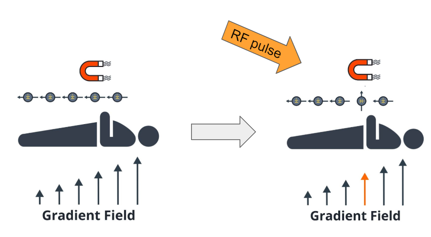
When an external radiofrequency pulse is applied, of a frequency proportional to the frequency of precession, the protons respond to this pulse in unison, or resonate, and flip the orientation of their spins. Once this pulse is gone, they return to their original orientation (along the static magnetic field).
The way in which protons return to their original orientation is different and depends on the tissue type that protons are a part of.
Since many protons are returning to their original orientation at once, they generate electrical currents in the coils that are placed nearby. Due to the resonance effect these currents are not insignificant and can be measured - these measurements constitute the data about the tissue being studied which is collected by the MRI scanner.
Gradient fields are used to vary the static magnetic field, and thus precession frequency, spatially. This allows the MR scanner to isolate a part of the body (i.e. a slice) that is being imaging. Further gradient fields are used to isolate information coming from specific locations within a slice.
EM fields
Electromagnetic fields of an MR scanner
What is MR scanner measuring?